Knee
Knee Anatomy
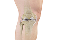
The knee is a complex joint made up of different structures including bones, tendons, ligaments and muscles. They all work together to maintain normal function and provide stability to the knee during movement.
Knee Arthritis
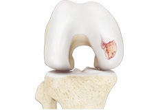
Arthritis is a general term covering numerous conditions where the joint surface or cartilage wears out. The joint surface is covered by a smooth articular surface that allows pain-free movement in the joint. This surface can wear out for several reasons; often the definite cause is not known.
Meniscus Tear
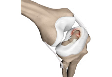
A meniscus tear is the commonest knee injury in athletes, especially those involved in contact sports. A sudden bend or twist in your knee can cause the meniscus to tear. This is a traumatic meniscal tear. Elderly people are more prone to degenerative meniscal tears as the cartilage wears out and weakens with age.
Articular Cartilage Defect
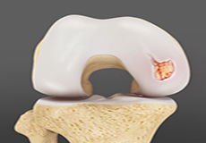
Articular or hyaline cartilage is the tissue lining the surface of the two bones in the knee joint. Cartilage helps the bones move smoothly against each other and can withstand the weight of the body during activities such as running and jumping.
Anterior Cruciate Ligament (ACL) Tear
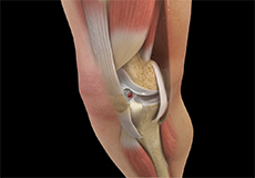
The anterior cruciate ligament, or ACL, is one of the major ligaments of the knee that is in the middle of the knee and runs from the femur (thighbone) to the tibia (shinbone). It prevents the tibia from sliding out in front of the femur. Together with posterior cruciate ligament (PCL) it provides rotational stability to the knee.
Patellofemoral Instability
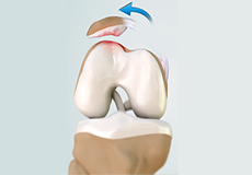
The knee can be divided into three compartments: patellofemoral, medial and lateral compartment. The patellofemoral compartment is the compartment in the front of the knee between the kneecap and thighbone. The medial compartment is the area on the inside portion of the knee, and the lateral compartment is the area on the outside portion of the knee joint.
Quadriceps Tendon Tear
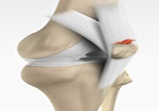
Quadriceps tendon is a thick tissue located at the top of the kneecap. The quadriceps tendon works together with the quadriceps muscles to allow us to straighten our leg. The quadriceps muscles are the muscles located in front of the thigh.
Osteonecrosis (AVN)
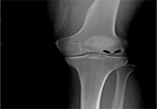
Osteonecrosis is a condition in which death of a section of bone occurs because of lack of blood supply to it. It is one of the most common causes of knee pain in older women. Women over 60 years of age are commonly affected, three times more often than men.
PCL Tear
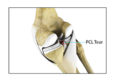
Coming soon
Posterolateral Instability

Coming soon
MCL Tear
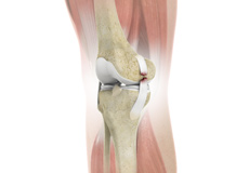
Coming soon
Osteochondral Defect (OCD)

Coming soon
ACL Reconstruction
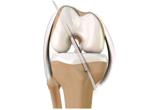
The anterior cruciate ligament is one of the major stabilizing ligaments in the knee. It is a strong rope like structure located in the centre of the knee running from the femur to the tibia.
Total Knee Replacement
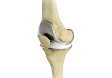
A Total Knee Replacement (TKR) or Total Knee Arthroplasty is a surgery that replaces an arthritic knee joint with artificial metal or plastic replacement parts called the 'prostheses'.
High Tibial Osteotomy
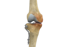
Knee osteotomy is a surgical procedure in which the upper shinbone (tibia) or lower thighbone (femur) is cut and realigned. It is usually performed in arthritic conditions affecting only one side of your knee and the aim is to take pressure off the damaged area and shift it to the other side of your knee with healthy cartilage.
Patellar Tendon Repair
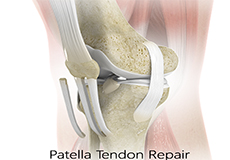
Patella tendon rupture is the rupture of the tendon that connects the patella (knee cap) to the top portion of the tibia (shin bone). The patellar tendon works together with the quadriceps muscle and the quadriceps tendon to allow your knee to straighten out.
Meniscal Surgery
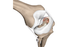
A meniscus tear is the commonest knee injury in athletes, especially those involved in contact sports. A sudden bend or twist in your knee can cause the meniscus to tear. This is a traumatic meniscal tear. Elderly people are more prone to degenerative meniscal tears as the cartilage wears out and weakens with age.
Patellofemoral Knee Replacement
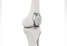
Traditionally, a patient with only one compartment of knee arthritis would undergo a total knee replacement surgery. Patellofemoral knee replacement is a minimally invasive surgical option that preserves the knee parts not damaged by arthritis as well as the stabilizing anterior and posterior cruciate ligaments, ACL and PCL.
Cartilage Repair and Transplantation
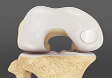
Articular cartilage is the white tissue lining the end of bones where these bones connect to form joints. Cartilage acts as cushioning material and helps in smooth gliding of bones during movement. An injury to the joint may damage this cartilage which cannot repair on its own.
Patella Stabilization

Patellar (kneecap) instability results from one or more dislocations or partial dislocations (subluxations).
Tibial Tubercle Osteotomy

Coming soon
MPFL Reconstruction
Coming soon
Patellafemoral Realignment

Coming soon
Distal Femoral Osteotomy (DFO)
Coming soon


